Blood Vessels Labeled Diagram - Blood Vessels | Free Blood Vessels Templates : We then simplified the anatomy of the heart even further with the below cartoon diagram and 2x2 table.. The inner surface of every blood vessel is lined by a thin layer of cells known as the endothelium. Once blood is oxygenated in the lungs, it returns to the heart and is then pumped throughout the body. Capillaries surround body cells and tissues to deliver and absorb oxygen, nutrients, and other substances. (ii) name the blood vessel supplying blood to the walls of the heart with oxygen. Arteries carry blood away from the heart to other organs.
(ii) name the blood vessel supplying blood to the walls of the heart with oxygen. It is through the capillaries that oxygen, nutrients, and wastes are exchanged between the blood and the tissues.the capillary networks are the ultimate destination of arterial blood from the heart and are the starting point for flow of venous blood back to the heart. Start studying blood vessels labeling. The sclera and cornea (opaque and transparent layer respectively) the choroid (filled with blood vessels) There are three major types of blood vessels:
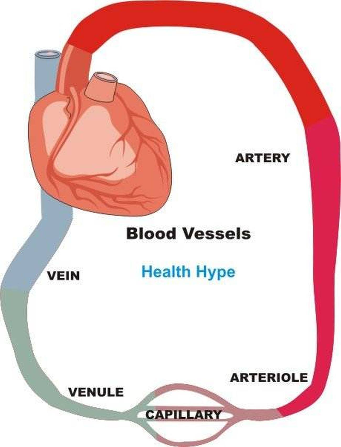
Pictures Of Blood Vessels | Healthiack from healthiack.com There are five main types of blood vessels: Use key choices to identify the blood vessel tunic described. Which labeled blood vessel shown in the diagram is the right common carotid artery? A blood vessel's main function is to transport blood around the body. Find the perfect blood vessel diagram stock photo. This blood is rich in oxygen. Arteries, arterioles, capillaries, venules and veins. Coronary vessels anatomical health care vector illustration labeled diagram.
Bulky middle tunic contains smooth muscle and elastin 3.
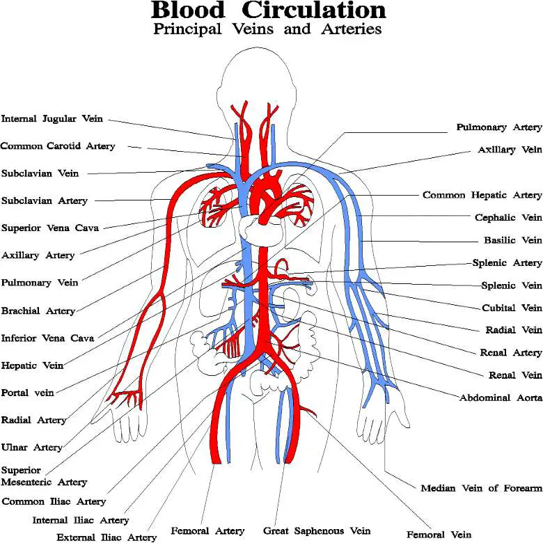
(ii) name the blood vessel supplying blood to the walls of the heart with oxygen. Learn vocabulary, terms, and more with flashcards, games, and other study tools. Use key choices to identify the blood vessel tunic described. Labeled diagram showing the structure of a blood vessel observe the blood vessels diagrams above, where you can see the structures of arteries and veins clearly labeled. Arteries and arterioles have thicker walls than veins and venules because they are closer to the heart and receive blood that is surging at a far greater pressure (figure 2). Blood vessels also play a role in controlling your blood pressure. Heart blood flow system with blood vessel scheme. Which labeled blood vessel shown in the diagram is the right common carotid artery? Start studying blood vessels labeling. The iliac, femoral, popliteal and tibial (calf) veins are the deep veins in the legs. A web of blood vessels—arteries, veins, and capillaries—circulate blood to organs. The 4 valves are the aortic, pulmonary, mitral, and tricuspid valves. Deep veins, located in the center of the leg near the leg bones, are enclosed by muscle.
Find the perfect blood vessel diagram stock photo. The eyeball is filled with vitreous humor, with the aqueous humor lying in the small anterior chamber of the eye. Use key choices to identify the blood vessel tunic described. Labeled diagram showing the structure of a blood vessel observe the blood vessels diagrams above, where you can see the structures of arteries and veins clearly labeled. Blood vessel structure & function from www.scritub.com which of the labeled layers in the diagram of the arterial wall is composed of a simple squamous epithelium, a basement membrane and a layer of.
# 72 Arteries, veins and capillaries - structure and functions | Biology Notes for IGCSE 2014 from 3.bp.blogspot.com The capillaries connect the two types of blood vessel and molecules are exchanged between the blood and. A blood vessel's main function is to transport blood around the body. Labeled diagram showing the structure of a blood vessel observe the blood vessels diagrams above, where you can see the structures of arteries and veins clearly labeled. Blood vessels are the specially designed tubes that carry blood throughout the body. Blood vessel structure & function from www.scritub.com which of the labeled layers in the diagram of the arterial wall is composed of a simple squamous epithelium, a basement membrane and a layer of. Which labeled blood vessel shown in the diagram is the right common carotid artery? Bulky middle tunic contains smooth muscle and elastin 3. Capillary, in human physiology, any of the minute blood vessels that form networks throughout the bodily tissues;
Use key choices to identify the blood vessel tunic described.
Blood vessels are the specially designed tubes that carry blood throughout the body. Learn even faster with this blood vessel anatomy study guide. Bulky middle tunic contains smooth muscle and elastin 3. Blood vessels are found throughout the body. Find the perfect blood vessel diagram stock photo. The capillaries connect the two types of blood vessel and molecules are exchanged between the blood and. The diagram below shows the human circulatory system. Labeled diagram showing the structure of a blood vessel observe the blood vessels diagrams above, where you can see the structures of arteries and veins clearly labeled. (ii) name the blood vessel supplying blood to the walls of the heart with oxygen. Arteries (in red) are the blood vessels that deliver blood to the body. Anatomy of the heart and main cardiac structures including the heart valves, chambers (atria and ventricles), and great vessels. Use key choices to identify the blood vessel tunic described. Which blood vessel shown in the diagram is the left subclavian artery?
Different types of blood vessels vary slightly in their structures, but they share the same general features. (ii) name the blood vessel supplying blood to the walls of the heart with oxygen. Which labeled blood vessel shown in the diagram is the right common carotid artery? Function and anatomy of the heart made easy using labeled diagrams of cardiac structures and blood flow through the atria, ventricles, valves, aorta, pulmonary arteries veins, superior inferior vena cava, and chambers. The left atrium receives blood from the lungs.
Blood vessels diagram from healthiack.com Which labeled blood vessel shown in the diagram is the right common carotid artery? The inner surface of every blood vessel is lined by a thin layer of cells known as the endothelium. Includes an exercise, review worksheet, quiz, and model drawing of an anterior vi Labeled diagram showing the structure of a blood vessel observe the blood vessels diagrams above, where you can see the structures of arteries and veins clearly labeled. Blood vessels are found throughout the body. We then simplified the anatomy of the heart even further with the below cartoon diagram and 2x2 table. These vessels connect other organs in your body to your heart. The capillaries connect the two types of blood vessel and molecules are exchanged between the blood and.
Learn even faster with this blood vessel anatomy study guide.
Heart blood flow system with blood vessel scheme. The cardiovascular system consists of the heart, blood vessels, and the approximately 5 liters of blood that the blood vessels transport. Once blood is oxygenated in the lungs, it returns to the heart and is then pumped throughout the body. Includes an exercise, review worksheet, quiz, and model drawing of an anterior vi Arteries carry blood away from the heart to other organs. Anatomy of the heart and main cardiac structures including the heart valves, chambers (atria and ventricles), and great vessels. We then simplified the anatomy of the heart even further with the below cartoon diagram and 2x2 table. It is returned to the heart in the veins. He has been with healthiack.com since 2012 and has written and reviewed well over 500 coherent articles. No need to register, buy now! A blood vessel's main function is to transport blood around the body. Different types of blood vessels vary slightly in their structures, but they share the same general features. The inner surface of every blood vessel is lined by a thin layer of cells known as the endothelium.
The sclera and cornea (opaque and transparent layer respectively) the choroid (filled with blood vessels) blood vessels labeled. This blood is rich in oxygen.
Blood Vessels Labeled Diagram - Blood Vessels | Free Blood Vessels Templates : We then simplified the anatomy of the heart even further with the below cartoon diagram and 2x2 table.. There are any Blood Vessels Labeled Diagram - Blood Vessels | Free Blood Vessels Templates : We then simplified the anatomy of the heart even further with the below cartoon diagram and 2x2 table. in here.